|
Biological Magnetic Resonance Data BankA Repository for Data from NMR Spectroscopy on Proteins, Peptides, Nucleic Acids, and other Biomolecules |
Member of
|
Mechanism: Hen's Egg White Lysozyme |
|||||
| |||||
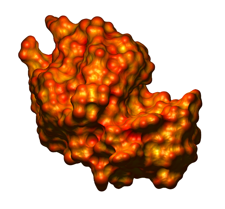
As stated previously, Lysozyme's active site is a long, deep cleft in the protein surface. This cleft binds a polysaccharide six amino sugars long (e.g. NAM-NAG-NAM-NAG-NAM-NAG). The polysaccharide substrate is properly positioned by hydrogen bonding and hydrophobic interactions. However, it isn't a perfect fit. In order for the 4th sugar unit to fit into its position, it must be distorted to form a less stable conformation. This increases the strain on the bond between the 4th and 5th sugar units, the bond that is to be broken. Certain amino acids present in the active site, Asp-52 and Glu-35, are vital for the catalytic action of Lysozyme. These residues interact with the 4th and 5th sugar units, breaking the carbon-oxygen bond between them by inserting a water molecule at this location (hydrolysis). First, the carboxylic acid sidechain of Glu-35 donates a proton to the oxygen of the unstable C-O bond, breaking it. This forms an intermediate with a positively charged (unstable) carbon atom. Asp-52 acts to stabilize this intermediate either through electrostatic interactions or by covalently bonding to the carbon. Then, an -OH from water attacks the carbon to yield the hydrolyzed end product. The extra H+ from water attacks the COO- of Glu-35 replenishing the proton that was lost earlier. Lysozyme then releases the broken chain and is able to bind a new substrate on the bacterial cell wall. Previous: Introduction |
